Since the necropsy facility at the park is 4WD-access-only, we caught a lift up with the vet (awesome guy, very generous with the beer & pizza) and his assistants. Truly, this is the best way to see a safari park, appreciate the rhino deciding not to park in the road, and get drooled on by a giraffe.

Left to right: Kristin (pathologist), Johanna (clinical path resident), me.
One never quite knows the appropriate expression to assume when posing with an ex-hippo...

The pale pink markings on the skin visible in the above and below pictures are scarring from frostbite when they had a really cold year and the pond froze. That was when they lost the previous hippo - she dived under the ice and drowned.

Hippo feet are cool.

Happiness is learning that a box-cutter is a perfect tool for getting through hippo skin.

The most time-consuming and exhausting part of the necropsy was simply reflecting the skin. There were multiple, often symmetrical subcutaneous haemorrhages (man, that felt awesome spelling that word with an 'a' again), I'm pointing at one with the knife:

Here is where we realise that pretty much everything in the cranial abdomen is in fact fused with the peritoneum... Most confusing when you're expecting to be able to get into the abdomen and find a pile of organs and suddenly there's spleen but no abdominal cavity....

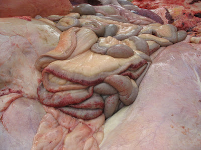
There were extensive areas of petechial haemorrhage on the serosal surface of the small and large intestines - the mucosal surface was even more extensively affected.

Here you can see the three large chambers of the fore-stomach, and an area of inflammation on the large intestine & mesentery that was overlying the diseased bladder.

The tip of the bladder is necrotic, and although it hadn't perforated it wouldn't have been far away...

Here's the opened bladder - at the right side is the necrotic end, a partially constricted sac filled with a 1.2lb urolith, mostly "sand" with some firm stones. Amazingly, this hippo hadn't actually blocked. We're not sure if the constriction in the bladder is a reaction to the stone, or if it was a pre-existing structure that subsequently filled with urolith.

The heart was cool - check out the extensive trabeculae.

I'm not sure if more hippo pics will turn up later - there were several cameras sporadically in use by whoever had "clean" hands.
3 comments:
so why was the cause of death specifically?
Septicaemia is most likely - supported by the haemorrhages everywhere.
I would never have thought I'd read about a Hippo's insides before.. interesting & informative!
Post a Comment